Полная версия
Common Objects of the Microscope
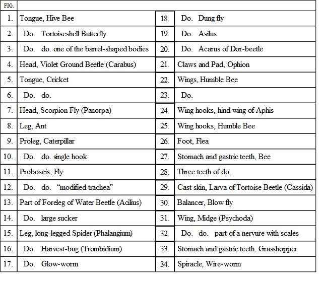
Fig. 1 on the same Plate shows the jaws of the hive bee, where the same organs are seen modified into many curious shapes. In the centre may be seen the tongue, elongated into a flexible and hair-covered instrument, used for licking the honey from the interior of flowers. At each side of the tongue are the labial palpi, having their outermost joints very small, and the others extremely large, the latter acting as a kind of sheath for the tongue. Outside the labial palpi are the maxillæ, separated in the specimen, but capable of being laid closely upon each other, and outside all are the mandibles.
VI.

VI.
The curiously elongated head of the scorpion-fly (Panorpa), seen at Fig. 7, affords another example of the remarkable manner in which these organs are developed in different insects. Another elongated head, belonging to the daddy long-legs, is seen in Plate VI. Fig. 27, and well shows the compound eyes, the antennæ, and the palpi. Fig. 2 represents the coiled tongue of the Atalanta butterfly; it is composed of the maxillæ, very greatly developed, and appearing as if each had originally been flat, and then rolled up so as to make about three-fourths of a tube. A number of projections are seen towards the tip, and one of these little bodies is shown on a larger scale at Fig. 3. These curious organs have probably some connection with the sense of taste. Along the edges of the semi-tubes are arranged a number of very tiny hooks, by means of which the insect can unite the edges at will.
Fig. 11, in the centre of the Plate, shows one of the most curious examples of insect structure, the proboscis or trunk of the common bluebottle-fly. The maxillary palpi covered with bristles are seen projecting at each side, and upon the centre are three lancet-like appendages, two small and one large, which are used for perforating various substances on which the insect feeds. The great double disc at the end is composed of the lower lip greatly developed, and is filled with a most complex arrangement of sucking-tubes, in order to enable it to fulfil its proper functions. The numerous tubes which radiate towards the circumference are strengthened by a vast number of partial rings of strong filamentary substance, like that which we have already seen in the breathing-tube of the silkworm. Some of these partial rings are seen on Fig. 12, a little above. The mode in which the horny matter composing the rings is arranged upon the tubes is most wonderful, and requires a tolerably high power to show it. The fine hairs upon the proboscis itself afford most admirable practice for the young microscopist. They should, when properly lighted and focused, be quite black and sharp. Any errors of manipulation will cause them to be “fuzzy.”
Fig. 5 shows the tongue of the common cricket, a most elegantly formed organ, having a number of radiating bands covered with zigzag lines, due to the triangular plates of strengthening substance with which they are furnished, instead of the rings. A portion more highly magnified is shown at Fig. 6, exhibiting the manner in which the branches are arranged.
The legs of insects now claim our attention.
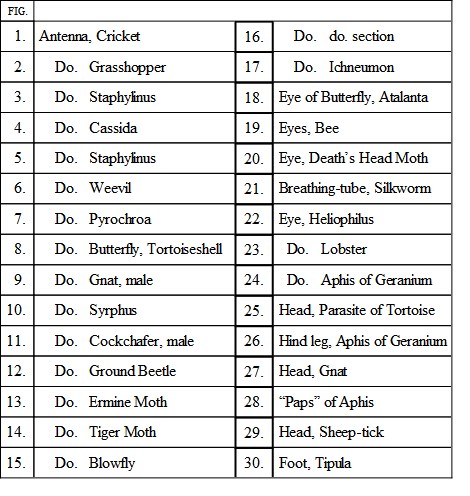
Fig. 9, Plate VII., shows the “pro-leg” of a caterpillar. The pro-legs are situated on the hinder parts of the caterpillar, and, being set in pairs, take a wonderfully firm hold of a branch or twig by pressure toward each other. Around the pro-legs are arranged a series of sharp hooks, set with their points inwards, for greater power in holding. Fig. 10 represents one of the hooks more magnified.
Fig. 15 is the lower portion of the many-jointed legs of the long-legged spider (Phalángium), the whole structure looking very like the antenna of the cricket. Fig. 17 is the leg of the glow-worm, showing the single claw with which it is armed. Fig. 26 shows the foot of the flea, furnished with two simple claws. Fig. 16 is the foot of the Trombídium, a genus of parasitic creatures, to which the well-known harvest-bug belongs. Fig. 26, Plate VI., shows the leg of the green Aphis of the geranium, exhibiting the double claw, and the pad or cushion, which probably serves the same purpose as the pads found upon the feet of many other insects. Fig. 8 is the lower portion of the leg of the ant, showing the two claws and the curious pad in the centre, by means of which the insect is able to walk upon slippery surfaces. The Típula has a foot also furnished with a single pad (see Plate VI. Fig. 30). This organ is seen under a very high power to be covered with long hair-like appendages, each having a little disc at the end, and probably secreting some glutinous fluid which will enable the creature to hold on to perpendicular and smooth surfaces. Many of my readers will doubtless have noticed the common fly, towards the end of autumn, walking stiffly upon the walls, and evidently detaching each foot with great difficulty, age and infirmity having made the insect unable to lift its feet with the requisite force.
Fig. 21 is the foot of one of the ichneumon-flies (Ophíon), the hairy fringe being apparently for the purpose of enabling it to hold firmly to the caterpillar in which it is depositing its eggs, and which wriggles so violently under the infliction that it would soon throw its tormentor had not some special means been provided for the purpose of enabling the latter to keep its hold. Fig. 20 is a beautiful example of a padded foot, taken from the little red parasitic creature so plentifully found upon the dor or dung beetle (Geotrúpes), and of which the afflicted insect is said to rid itself by lying on its back near an ant’s nest, and waiting until the ants carry off its tormentors.
Fig. 18 is the foot of the common yellow dung-fly (plentiful in pasture lands), having two claws and two pads; and Fig. 19 shows the three pads and two claws found in the foot of the hornet-fly (Ásilus).
Few microscopic objects call forth such general and deserved admiration as the fore-foot of the male water-beetle (Dytiscus), when properly prepared and mounted, for which see Fig. 13.
On examining this preparation under the microscope, it is seen that three of the joints are greatly expanded, and that the whole of their under surface is covered profusely with certain wonderful projections, which are known to act as suckers. One of them is exceedingly large, and occupies a very considerable space, its hairs radiating like the rays of the heraldic sun. Another is also large, but scarcely half the diameter of the former, and the remainder are small, and mounted on the extremities of delicate foot-stalks, looking something like wide-mouthed trumpets. In the specimen from which the drawing was taken the smaller suckers are well shown, as they protrude from the margin of the foot.
One of the larger suckers is seen more magnified on Fig. 14.
Plate VIII. Fig. 1, exemplifies the manner in which the muscles of insects do their work, being well attached in the limbs to the central tendon, and pulling “with a will” in one direction, thus giving very great strength. This leg is taken from the water boatman (Notonecta), and has been mounted in Canada balsam.
On Plate VII. Fig. 29, may be seen a curiously formed creature. This is the larva of the tortoise beetle (Cássida), the skin having been flattened and mounted in Canada balsam. The spiracles are visible along the sides, and at the end is seen a dark fork-like structure. This is one of the peculiarities of this creature, and is employed for the purpose of carrying the refuse of its food, which is always piled upon its back, and retained in its place by the forked spines, aided probably by the numerous smaller spines that project from the side.
Fig. 33 shows part of the stomach and gastric teeth of the grasshopper. This structure may be seen to perfection in the “gizzard,” as it is called, of the great green locust of England (Ácrida viridíssima). The organ looks like a sudden swelling of the œsophagus, and when slit longitudinally under water, the teeth may be seen in rows set side by side, and evidently having a great grinding power. The common house cricket has a similar organ of remarkable beauty. Just above (Fig. 27) is the corresponding structure in the hive bee, three of the teeth being shown separately at Fig. 28.
We now cast a rapid glance at the wings of insects.
They have no analogy, except in their use, with the wings of birds, as they are not modifications of existing limbs, but entirely separate organs. They consist of two membranes united at their edges, and traversed and supported by sundry hollow branches or “nervures,” which admit air, and serve as useful guides to entomologists for separating the insects into their genera. Indeed, the general character of the wings has long been employed as the means of dividing the insect race into their different orders, as may be seen in any work on entomology. The typical number of wings is four, but it often happens that two are almost wholly absent, or that the uppermost pair are thickened into a shelly kind of substance which renders them useless for flight; while in many insects, such as the ground beetles and others, the upper wings become hardened into firm coverings for the body, and the lower pair are shrivelled and useless.
Fig. 22 shows two of the wings of a humble bee, together with their nervures, and the peculiar system by which the upper and lower pair are united together at the will of the insect. At the upper edge of the lower wing, and nearly at its extremity, may be seen a row of very tiny hooks, shown on a larger scale at Fig. 25. These hooklets hitch into the strengthened membrane of the upper wing, which is seen immediately above them, and so conjoin the two together. The curious wing-hooks of the Aphis may be seen on Fig. 24, very highly magnified.
Fig. 31 is the wing of the midge (Psychóda), that odd little insect which is seen hopping and popping about on the windows of outhouses and similar localities, and is so hard to catch. The whole wing is plentifully covered with elongated scales, and is a most lovely object under any power of the microscope. These scales run along the nervures and edges of the wings, and part of a nervure is shown more highly magnified at Fig. 32.
At Fig. 23 is shown the wing of one of the hemipterous insects, common along the banks of ditches and in shady lanes, and known by the name of Cíxius. It is remarkable for the numerous spots which stud the nervures, one being always found at each forking, and the others being very irregularly disposed.
Fig. 30 is one of the balancers or “haltéres” of the house-fly. These organs are found in all the two-winged insects, and are evidently modifications of the second pair of wings. They are covered with little vesicles, and protected at their base by scales. Some writers suppose that the sense of smell resides in these organs. Whatever other purpose they may serve, they clearly aid in the flight, as, if the insect be deprived of one or both of the balancers, it has the greatest difficulty in steering itself through the air.
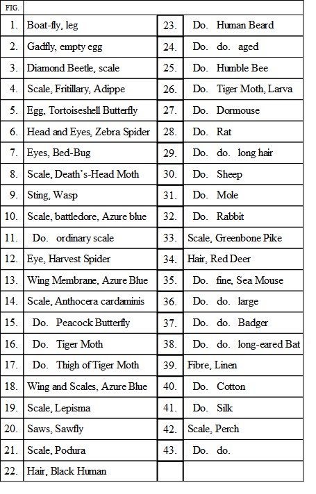
The wings of insects are mostly covered with hairs or scales, several examples of which are given in Plate VIII. Fig. 4 shows one of the scales of the Adippe or fritillary butterfly, exhibiting the double membrane—part of which has been torn away—and the beautiful lines of dots with which it is marked. The structure of the scales is further shown by a torn specimen of tiger moth scale seen on Fig. 16. On many scales these dots assume a “watered” aspect when the focus or illumination changes, an example of which may be seen in Fig. 15, a scale of the peacock butterfly.
Fig. 11 is one of the ordinary scales of the azure blue butterfly, and Fig. 10 shows one of the curious “battledore” scales of the same insect, with its rows of distinct dottings. Fig. 14 is one of the prettily tufted scales of the orange-tip butterfly, and Fig. 8 is the splendid branched scale of the death’s-head moth. Fig. 19 shows a scale of the sugar-runner (Lepisma saccharína), a little silvery creature with glistening skin, and long bristles at the head and tail, that is found running about cupboards, window-sills, and similar places. It is not easy to catch with the fingers, as it slips through them like oil; but by holding a cover-glass in a pair of forceps, and pressing it upon one of the little creatures, a number of the scales may be caused to adhere to it, and these should be mounted dry for examination. The gnats also possess very pretty scales, with the ribs projecting beyond the membrane.
VII.

VII.
Fig. 21 is a scale from the common spring-tail (Podúra plúmbea), a little creature which is found plentifully in cellars and other damp places, skipping about with great activity. Some flour scattered on a piece of paper is a sure trap for these little beings. Fig. 3 is one of the scales taken from the back of the celebrated diamond beetle, showing the cause of the magnificent gem-like aspect of that insect. We have in England many beetles of the same family—the weevils—which, although much smaller, are quite as splendid when exhibited under a microscope by reflected light. The wing-case or “elytron” of a little green weevil, very common in the hedges, may be seen on Plate XII. Fig. 10.
The reader will observe that all these scales are furnished with little root-like appendages, by means of which they are affixed to the insect. Fig. 13 shows a portion of the wing of the azure blue butterfly, from which nearly all the scales have been removed, for the purpose of exhibiting the pits or depressions in which they had formerly been fastened, and one or two of the scales are left still adherent to their places. The scales are arranged in equal rows like the slates of a housetop, as may be seen on Fig. 18, which represents part of the same wing, to show the scales overlapping each other, and the elegant form which they take near the edges of the wing, so as to form a delicate fringe. The long hair-like down which covers the legs and bodies of the moths and butterflies (which are called Lepidóptera, or scale-winged insects, in consequence of this peculiarity), is seen under the microscope to be composed of scales very much elongated, as is shown in Fig. 17, a portion taken from the leg of a tiger moth.
The eggs of insects are all very beautiful, and three of the most curious forms are given on Plate VIII.
Fig. 2 is the empty egg of the gad-fly, as it appears when fastened to a hair of the horse. Fig. 5 represents the pretty ribbed egg of the common tortoise-shell butterfly; and Fig. 7 is the very beautiful egg of the very horrid bed-bug, worthy of notice on account of the curious lid with which its extremity is closed, by means of which the young larva creeps out as soon as it is hatched.
The feathers of birds, and the fur of animals, will furnish many examples of the eggs of parasites, some of which are of extreme beauty. The feather or hair may be mounted in a cell without disturbing the eggs, which should, however, be heated sufficiently to kill the embryo if present.
Fig. 9 shows the penetrating portions of the sting of the wasp. The two barbed stings, which seem to be the minute prototypes of the many-barbed spears of the South Sea islanders, are seen lying one at each side of their sheath, and a single barb is drawn a little to the left on a very much larger scale. It is by reason of these barbs that the sting is always left adhering to the wound, and is generally drawn wholly out of the insect, causing its death in a short while.
The sting is only found in female insects, and is supposed to be analogous to the “ovipositor” of other insects, i.e. the instrument by which the eggs are deposited in their places. Fig. 20 shows the curious egg-placing apparatus of one of the saw-flies. The backs of these “saws” work in grooves, and they work alternately, so that the fly takes but a very short time in cutting a slit in the young bark of a tender shoot, and laying her eggs in the slit. When she has completed one of these channels, she sets to work upon another, and in the early spring the young branches of the gooseberry bushes may be seen plentifully covered with these grooves and the eggs. When hatched, black caterpillar-like grubs from the eggs issue, and devastate the bushes sadly, turning in process of time into blackish flies, which are seen hovering in numbers over the gooseberries, and may be killed by thousands.
The scales and hairs of other animals deserve great attention. Fig. 23 is a single hair of the human beard, as it often appears when tied in a knot—by Queen Mab and her fairies, according to Mercutio. Fig. 22 is a portion of the same hair as it appears when splitting at its extremity. The structure of the hair is not, however, so well seen in this object as in that represented on Fig. 24, which is a beautiful example of white human hair that once adorned the head of the victor of Waterloo. It formed one of a tiny lock given to me by a friend, and is so admirable an example of human hair, that I forthwith mounted it for the microscope. In this hair the cells may be seen extending down its centre, and the peculiar roughened surface produced by the flattened cells which are arranged around its circumference are also seen. By steeping in caustic potash, these scales can be separated, but generally they lie along the hair in such a manner that if the hair be drawn through the fingers from base to point, their projecting ends permit it to pass freely; whilst if it be drawn in the reverse direction, they cause it to feel very harsh to the touch.
In the sheep’s wool (Fig. 30) this structure is much more developed, and gives to the fibres the “felting” power that causes them to interlace so firmly with each other, and enables cloth—when really made of wool—to be cut without unravelling. Fig. 37 is the smooth hair of the badger; and Fig. 34 is the curious hair of the red deer, which looks as if it had been covered with a delicate net.
Fig. 28 is the soft, grey, wool-like hair of the rat; and Fig. 29 is one of the larger hairs that protrude so plentifully, and form the glistening brown coat of that animal. Fig. 38 is the curiously knobbed hair of the long-eared bat, the knobs being formed of protuberant scales that can easily be scraped off. Fig. 31 shows a hair of the common mole; and Fig. 32 is one of the long hairs of the rabbit. Fig. 27 is a flat hair of the dormouse, slightly twisted, the difference in the breadth showing where the twist has taken place. The hair of the mouse is beautifully ribbed, so as to look like a ladder. Fig. 26 is one of the very long hairs that so thickly clothe the tiger moth caterpillar; and Fig. 25 is a beautifully branched hair taken from the common humble bee.
All hairs should be examined by polarised light, with a plate of selenite, when most gorgeous colour effects may be obtained.
The four fibres mostly used in the manufacture of apparel are: wool, Fig. 30, which has already been described; linen, Fig. 39; cotton, Fig. 40; and silk, Fig. 41. The structure of each is very well marked and easily made out with the microscope; so that an adulterated article can readily be detected by a practised eye. Cotton is the most common adulteration of silk and linen fabrics, and may at once be detected by its flat twisted fibre. Silk is always composed of two parallel threads, each proceeding from one of the spinnerets of the caterpillar, and it may be here remarked that if these threads are not quite parallel the silk is of bad quality. Silken fibre is always covered, when new, with a kind of varnish, usually of a bright orange colour, which gives the undressed “floss” silk its peculiar hue, but which is soluble and easily washed away in the course of manufacture.
Figs. 35 and 36 are the small and large hairs of that magnificent creature, the sea mouse (Aphrodíte aculeáta), whose covering, although it lies in the mud, glows with every hue of the rainbow, and in a brilliant light is almost painfully dazzling to the eye.
VIII.

VIII.
The scales of some of the fishes are shown on Plate VIII., in order to exhibit their mode of growth by successive layers. The scales are always enveloped in membranous sacs, and in some cases, as in the eel, they do not project beyond the surface, and require some little observation to detect them. A scale of an eel is shown on Plate XI. Fig. 14, and is a magnificent object under polarised light. Fig. 33 is a scale of the greenbone pike; and Figs. 42 and 43 are scales of the perch, showing the roots by which they are held in their places. The roach, dace, bleak, and many other similar fish have a beautiful silvery substance on the under surface of the scales, which was greatly used in the manufacture of artificial pearls, glass beads being thinly coated in the interior with the glittering substance, and then filled in with wax. A piece of sole-skin, when preserved in Canada balsam and placed under the microscope, is a very beautiful object.
More examples of hairs, and other processes from the skin, together with the structure of the skin itself, of bone, of blood, and the mode in which it circulates, are given on Plate X.
In all important points of their structure the feathers of birds are similar to the hairs of animals, and are developed in a similar manner. They are all composed of a quill portion, in which the pith is contained, and of a shaft, which carries the vane, together with its barbs. The form of each of these portions varies much, even in different parts of the same bird, and the same feather has almost always two kinds of barbs; one close and firm, and the other loose, floating, and downy. If a small feather be plucked from the breast or back of a sparrow or any other small bird, the upper part of the feather is seen to be close and firm, while the lower is loose and downy, the upper part being evidently intended to lie closely on the body and keep out the wet, while the lower portion affords a soft and warm protection to the skin.
Fig. 12, Plate X., shows the feather of a peacock, wherein the barbs are very slightly fringed and lie quite loosely side by side. Fig. 18 is part of the same structure, in a duck’s feather, wherein are seen the curious hooks which enable each vane to take a firm hold of its neighbour, the whole feather being thus rendered firm, compact, and capable of repelling water. The reader will not fail to notice the remarkable analogy between these hooks and those which connect the wings of the bee.
Fig. 17 is a part of the shaft of a young feather taken from the canary, given for the purpose of showing the form of the cells of which the pith is composed. Fig. 20 is part of the down from a sparrow’s feather, showing its peculiar structure; and Fig. 21 is a portion of one of the long drooping feathers of the cock’s tail.
Fig. 13 exhibits a transverse section of one of the large hairs or spines from the hedgehog, and shows the disposition of the firm, horn-like exterior, and the arrangement of the cells. Sections of various kinds of hair are interesting objects, and are easily made by tying a bundle of them together, soaking them in gum, hardening in spirit, and then cutting thin slices with a razor. A little glycerine will dissolve the gum, and the sections of hair will be well shown. Unless some such precaution be taken, the elasticity of the hair will cause the tiny sections to fly in all directions, and there will be no hope of recovering them.
Several examples of the skin are also given. Fig. 27 is a section through the skin of the human finger, including the whole of one of the little ridges which are seen upon the extremity of every finger, and half of two others. The cuticle, epidermis, or scarf-skin, as it is indifferently termed, is formed of cells or scales, much flattened and horny in the upper layers, rounder and plumper below. The true skin, or “cutis,” is fibrous in structure, and lies immediately beneath, the two together constituting the skin, properly so called. Beneath lies a layer of tissue filled with fatty globules, and containing the glands by which the perspiration is secreted.